Gastrulation and development of organ systems
The CNS system involves 3 germinal layers: ectoderm, mesoderm, and endoderm. The ectoderm is the key initiating player in the embryogenesis of the CNS. The ectoderm is further sub-specialized as the (1) surface ectoderm, which differentiates into the epidermis, nails, and hair. The ectoderm is also sub-specialized to form the (2) neural.

Major Structures Arising Out Of Primary Germ Layers Embryogenesis MCAT Content
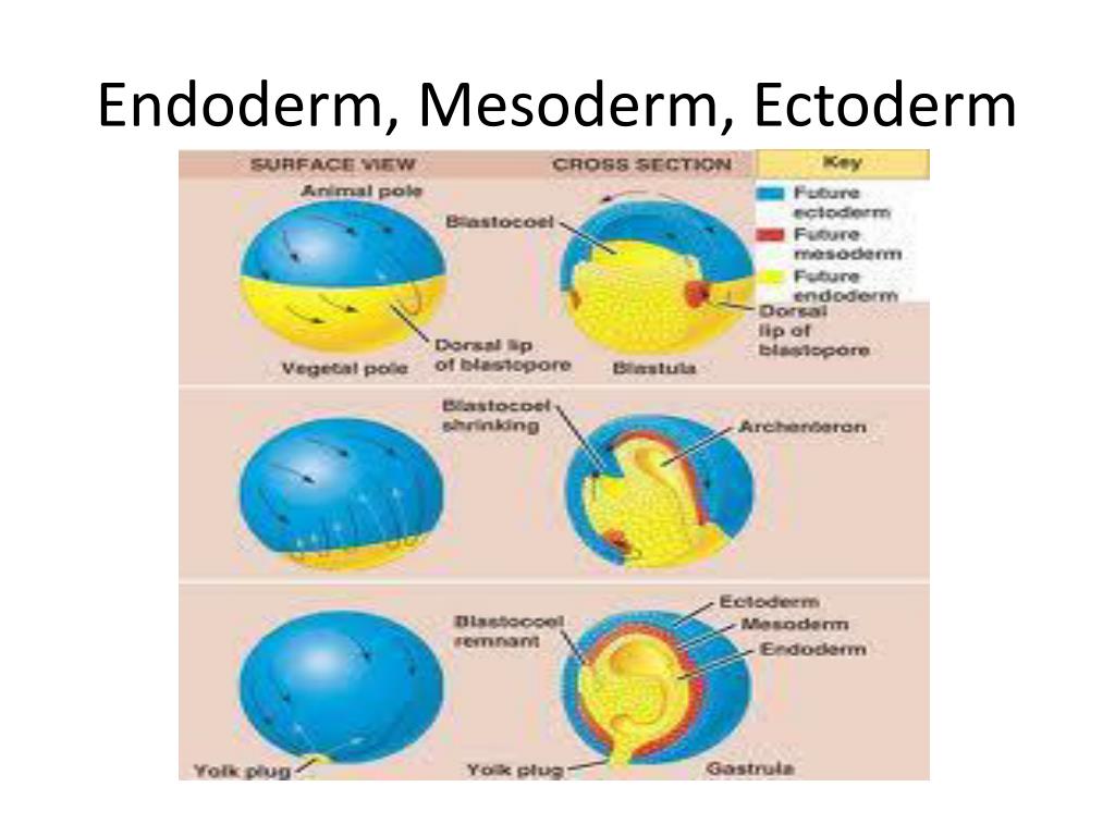
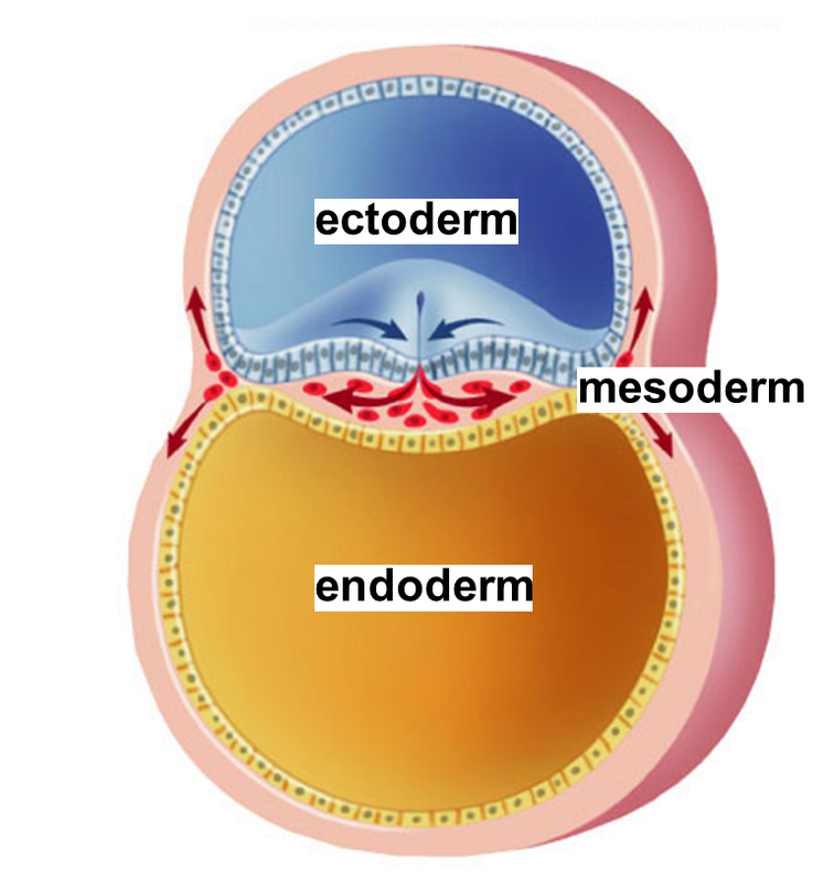
Gastrulation is a phase that occurs during the third week of early embryonic development, and it takes place immediately after the blastula phase in most animals. Here, the single-layered and hollow blastula is rearranged and differentiated in the multi-layered gastrula with three distinct layers: the ectoderm, mesoderm, and endoderm.

SoftwarePhysics May 2014
Mesoderm, the middle of the three germ layers, or masses of cells (lying between the ectoderm and endoderm), which appears early in the development of an animal embryo. In vertebrates it subsequently gives rise to muscle, connective tissue, cartilage, bone, notochord, blood, bone marrow, lymphoid

28.2 Embryonic Development Anatomy & Physiology
In human embryology, weeks six through eight are characterized by the growth and differentiation of tissues into organs. This process is known as organogenesis and occurs from weeks three through eight; the embryonic period. During week three, the process of gastrulation occurs, which establishes three distinct cell layers; the mesoderm, endoderm, and ectoderm. These are the primary germ cell.
Introduction to ectoderm
The term endoderm is sometimes used to refer to the gastrodermis, the simple tissue that lines the digestive cavity of cnidarians and ctenophores. Compare ectoderm; mesoderm. Endoderm, the innermost of the three germ layers, or masses of cells (lying within ectoderm and mesoderm), which appears early in the development of an animal embryo.
PPT 4 PowerPoint Presentation, free download ID3070951
Abstract. The three germ layers — mesoderm, endoderm and ectoderm — constituting the cellular blueprint for the tissues and organs that will form during embryonic development, are specified at gastrulation. Cells of mesodermal origin are the most abundant in the human body, representing a great variety of cell types, including the.
PPT Organismal Development Part 5 PowerPoint Presentation, free download ID2368275
AboutTranscript. Explore the formation of specific structures from the three primary germ layers - endoderm, mesoderm, and ectoderm - during early embryogenesis. Discover how endoderm cells shape our gastrointestinal and pulmonary systems, mesoderm forms muscles, skeletal system, and genitourinary tracts, and ectoderm creates skin, related.

SOLUTION Anatomy mesoderm ectoderm and endoderm Studypool
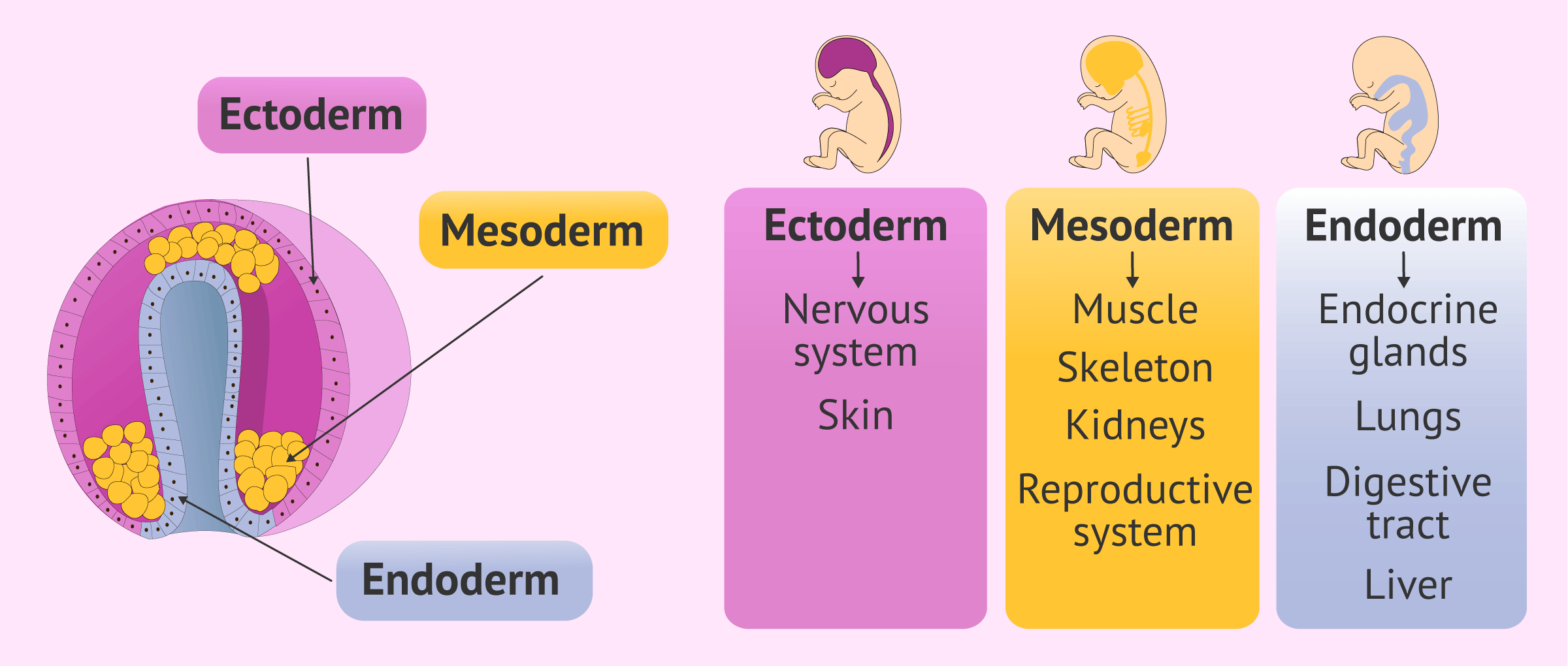
Topic: Embryogenesis. Ectoderm, mesoderm, and endoderm are the major structures arising out of the primary germ layers. Organogenesis is the phase of embryonic development that starts at the end of gastrulation and continues until birth. During organogenesis, the three germ layers formed from gastrulation: the ectoderm, endoderm, and mesoderm.
PPT Derivatives of the ectodermal germ layer PowerPoint Presentation ID1981616
The mesoderm germ layer forms in the embryos of triploblastic animals. During gastrulation, some of the cells migrating inward contribute to the mesoderm, an additional layer between the endoderm and the ectoderm. The formation of a mesoderm leads to the development of a coelom. Organs formed inside a coelom can freely move, grow, and develop.

BIOLOGY FORM 6 EMBRYOLOGY
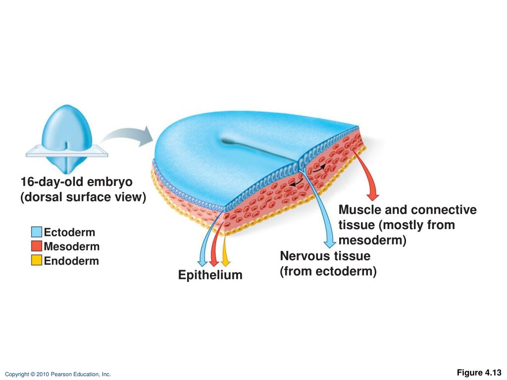
The entire nervous system forms via the process called neurulation in which neural tube and neural crest form initially. In the third week of embryogenesis, three germ layers arise, namely, ectoderm, mesoderm, and endoderm, through the process of gastrulation. The overlying ectoderm is induced and thickened by the notochord and the neural plate forms. The neural plate then gives rise to the.
Reproduction and Development AP biology
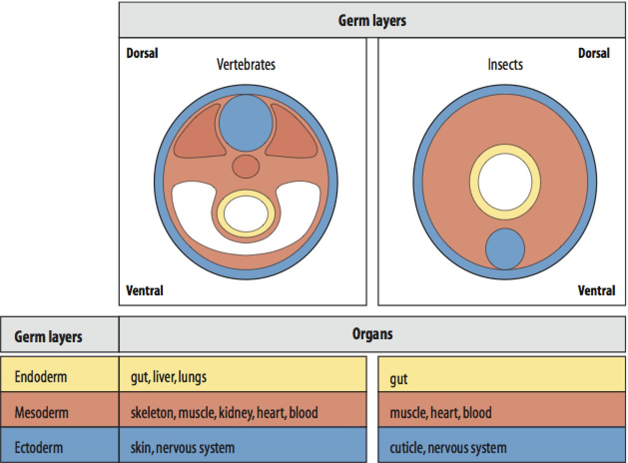
The gastrointestinal (GI) system involves three germinal layers: mesoderm, endoderm, ectoderm. Mesoderm gives rise to the connective tissue, including the wall of the gut tube and the smooth muscle. Endoderm is the source of the epithelial lining of the gastrointestinal tract, liver, gallbladder, pancreas.

Pin on біологія
It is formed during gastrulation, in which pluripotent epiblast cells are allocated to the three principal germ layers—ectoderm, mesoderm and definitive endoderm 1,2. The initiation of this.

How are the three germ layers formed?
germ layer, any of three primary cell layers, formed in the earliest stages of embryonic development, consisting of the endoderm (inner layer), the ectoderm (outer layer), and the mesoderm (middle layer). The germ layers form during the process of gastrulation, when the hollow ball of cells that constitutes the blastula begins to differentiate.

Ectoderm, Endoderm, & Mesoderm YouTube
Gastrulation leads to three germ layers—ectoderm, mesoderm and endoderm—that are separated by two basement membranes. In the mouse embryo, the emergent gut endoderm results from the widespread.

Mesoderm vector illustration VectorMine Nursing school notes, Human body anatomy, Physiology
The 3 germ layers - the ectoderm, the mesoderm, and the entoderm (endoderm): are in place at the end of gastrulation; THE ECTODERM gives rise to the central nervous system (the brain and spinal cord); the peripheral nervous system; the sensory epithelia of the eye, ear, and nose; the epidermis and its appendages (the nails and hair); the mammary glands; the hypophysis; the subcutaneous glands.

Formation of Mesoderm. Formation and segregation of the mesodermal germ... Download Scientific
Formation of the three primary germ layers, ectoderm, mesoderm, and endoderm, is an early distinction between groups of cells in developing embryos. Our understanding of their generation in vertebrates has benefitted from the classical experiments of Nieuwkoop and his colleagues (referenced in Nieuwkoop, 1997), in which explants of tissue from the animal hemisphere of amphibian embryos (fated.